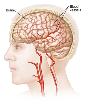
Understanding Cerebral Angiography
This test makes X-ray images of blood vessels in your brain. During the test, the healthcare provider puts a long, thin, flexible tube (catheter) into a blood vessel and moves it to the brain. They use a special contrast fluid to make the blood vessels in the brain show up on the X-rays. This test is also called arteriography.
Why cerebral angiography is done
This test is done to help find problems with the blood vessels in the brain. These may include:
-
Weakened area of blood vessel (aneurysm)
-
Tangle of arteries and veins (arteriovenous malformation or AVM)
-
Blood clots or bleeding. This may have caused a stroke or another problem.
-
Brain tumor
-
Inflammation of the blood vessels (vasculitis)
-
Evaluation of arteries before surgery
-
Stroke
Before you have this test, tell your provider if you:
-
Have bleeding problems
-
Take medicines that thin your blood such as warfarin or aspirin
-
Have allergies to X-ray contrast dye or iodine
-
May be pregnant
-
Have problems with your kidney function
How cerebral angiography is done
The test is done in a hospital or procedure center. You will likely go home the same day.
-
You lie on an exam table. Your head may be held still with straps or another device.
-
An IV (intravenous) line is put into a vein in your arm or hand. This provides fluids and medicines.
-
You may be given a medicine that helps you relax (sedative).
-
The staff will watch your heart activity with an ECG (electrocardiogram) machine. They will put sticky patches on your chest, arms, and legs and attach those with wires to the ECG machine.
-
The healthcare provider will prepare the site where the catheter will be inserted. The insertion site is usually in the groin. The site is cleaned. It's also numbed with an injection of anesthetic.
-
The healthcare provider makes a small incision or puncture into the artery at the insertion site. They put the catheter into the artery.
-
Using X-ray images as a guide, the healthcare provider carefully moves the catheter through the artery to the brain.
-
The healthcare provider injects contrast fluid through the catheter into the artery. You may feel warmth or pressure in your neck, face, or head.
-
The healthcare provider takes X-rays.
-
When the procedure is complete, the catheter is removed.
-
The staff will put pressure on the insertion site for a time to stop any bleeding.
-
When you are released to go home, have an adult family member or friend drive you.
Risks of cerebral angiography
© 2000-2024 The StayWell Company, LLC. All rights reserved. This information is not intended as a substitute for professional medical care. Always follow your healthcare professional's instructions.